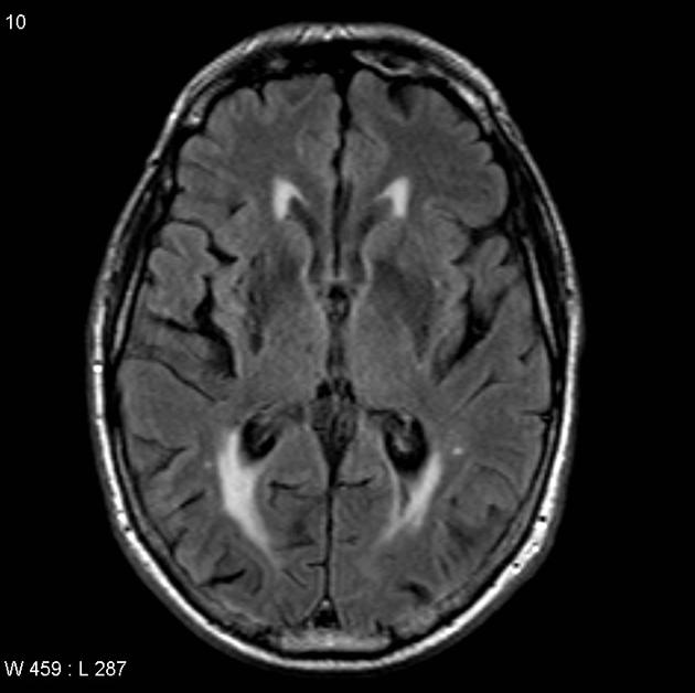
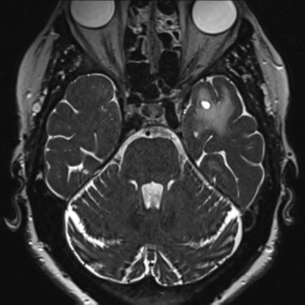
Dilated Perivascular Space

Enlarged perivascular spaces epvs or virchow robin spaces are cerebrospinal fluid filled cavities that surround small penetrating cerebral arterioles and correspond with extensions of the subarachnoid space.
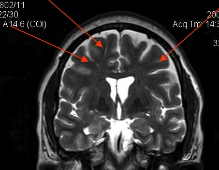
Dilated perivascular space. The mechanism that occurs is not well known but there are many hypotheses in study. Dilated perivascular spaces are principally places where the axons and the myelin sheath used to be. Recent animal and human studies have shown an increased frequency of enlarged high convexity virchow robin spaces vrs in several neurologic diseases suggesting their role as neuroradiologic markers of inflammatory changes.
Dilated perivascular spaces consist of regular cavities containing an artery. We believe that the enlargement of the perivascular space might reflect the accumulation of inflammatory cells and or changes of vascular permeability. Dilated vrs could provide a passive access to cns of blood borne cells which in turn could trigger or enhance the inflammatory process.
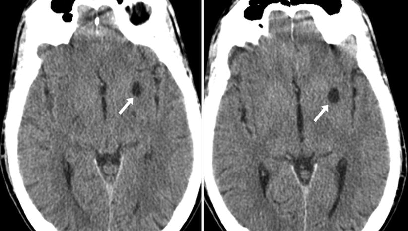
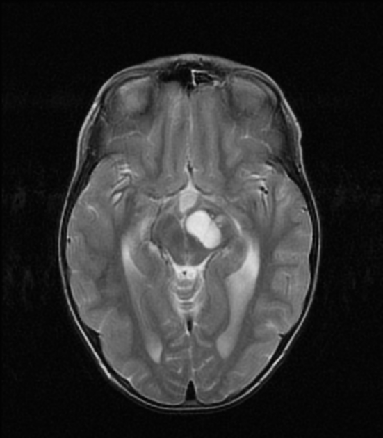
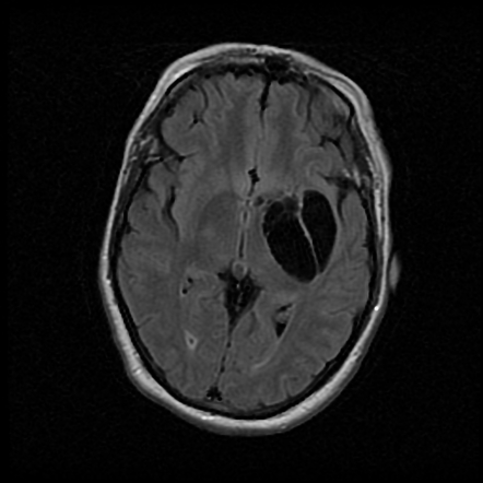
Dilated perivascular spaces have been shown to be a specific biomarker of cerebral small vessel disease in young patients with dementia. With the 1 5 t scanners concentrations of pathology as small as 2mm could be seen. Perivascular spaces which are visible on imaging are typically less than 5 mm in diameter but can reach much larger sizes.
In the brain perivascular cuffs are regions of leukocyte aggregation in the perivascular. They tend to enlarge with age and hypertension. What is being seen with dilated perivascular spaces is not damage to individual axons but damage to axonal tracts concentrated in a specific area.
1 epvs are visible on axial t2 weighted cerebral mri as characteristic small high signal areas in the basal ganglia and centrum semiovale that follow the orientation of penetrating arterioles. It is considered to be dilated when the size exceeds 2 mm visualized better in t2 weighted images. The brain pia mater is reflected from the surface of the brain onto the surface of blood vessels in the subarachnoid space.
The aim of this study was to determine the prevalence of high convexity dilated vrs in mild traumatic brain injury tbi. Our aim was to examine the discriminative power of dilated cerebral perivascular spaces as biomarkers of small vessel disease in a very elderly population of patients with dementia.
